FIG. 8.
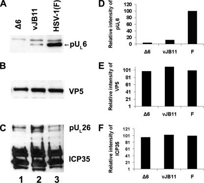
Immunoblot of B capsids probed with anti-pUL6, ICP35, or VP5 antibodies. Capsids were purified from CV1 cells infected with UL6 null virus, vJB11, or HSV-1(F) as detailed in Materials and Methods. B capsids denatured in SDS were either diluted 10-fold (B and C) or were loaded undiluted (A) onto an SDS-polyacrylamide gel through which they were separated by electrophoresis and transferred to a nitrocellulose sheet. The nitrocellulose sheet was probed with pUL6 (A), VP5 (B), or ICP35 (C) specific antibodies, and bound immunoglobulin was revealed by reaction with appropriately conjugated secondary antibodies followed by enhanced chemiluminescence. (D) The relative amounts of pUL6 in lanes 1 to 3 were determined using an LAS-3000 Mini Fujifilm imaging system. The chemiluminescent intensity of each pUL6-containing band in panel A is reported as a percentage of the signal obtained in panel A, lane 3. (E) Similar to panel D except that values reflect chemiluminescent intensities of VP5 in lanes of panel B normalized to that in panel B, lane 3. (F) Similar to panel D, but values reflect the ICP35 specific signals in panel C normalized to panel C, lane 3.

