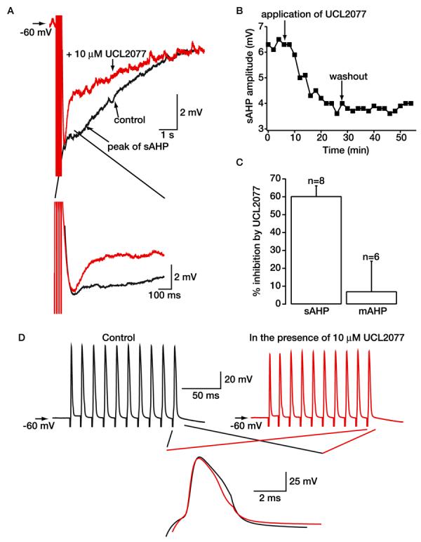
Fig. 4.

Effect of UCL2077 on the sAHP in hippocampal neurons present in the slice preparation. A, representative illustrations of the sAHP produced by an action potential train at −60 mV in hippocampal CA1 pyramidal neurons present in the slice preparation before and after application of 10 μM UCL2077. As explained in the text, concentrations of UCL2077 lower than 3 μM were ineffective in the slice preparation; presumably, as is known to occur with many compounds, it is sequestered by tissue. Note that UCL2077 had little effect on the mAHP (shown on an enhanced time scale in the inset). B, time course of the effects of 10 μM UCL2077 on the sAHP shown in A. C, bar graph to show the relative inhibition by 10 μM UCL2077 of the sAHP and mAHP recorded from hippocampal neurons present in the slice preparation. D, records of trains of action potentials in the absence and presence of 10 μM UCL2077. Superimposed traces of the last action potential under control conditions (black) and with UCL2077 present (red) are shown on an enhanced time scale.
