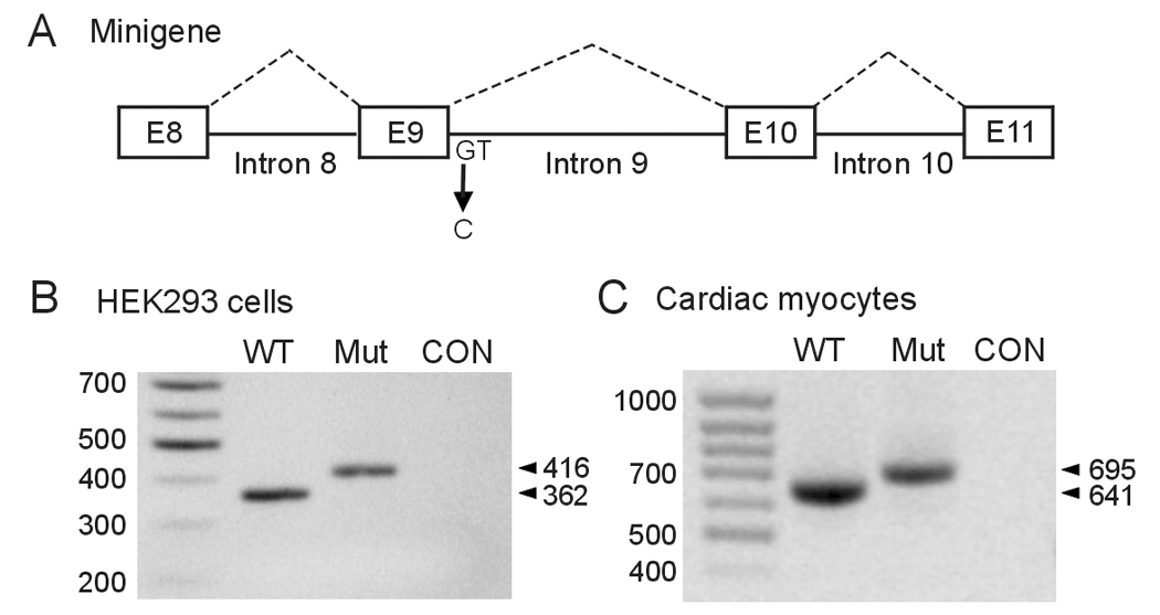
Figure 2.

Analysis of the 2398+1G>C mutation using minigenes expressed in HEK293 cells and neonatal rat ventricular myocytes. A: Diagram of the hERG minigene used in transfection experiments. B: The WT and 2398+1G>C minigenes were transfected into HEK293 cells and isolated RNAs were amplified by RT-PCR with the same primers as used in figure 1. CON: untransfected HEK293 cells. C: The WT and 2398+1G>C minigenes were infected into neonatal rat ventricular myocytes using adenovirus constructs. RT-PCR was performed using the same forward primer as above and a reverse primer complementary to the sequence in the recombinant adenovirus. CON: uninfected myocytes. Results shown are representative of three independent experiments.
