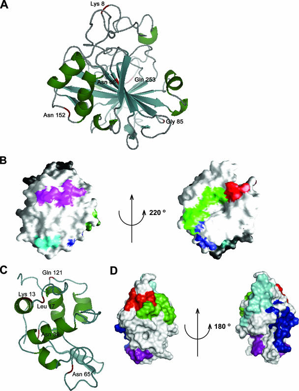
FIG. 6.
Surface residues of BCA are shown which interact with domain V RNA. (A) Ribbon diagram of BCA (PDB identifier 1V9E) showing five residues (labeled and marked in red) that interact with RNA during folding. Four of these amino acids are located in the loop regions. The cartoon was drawn using the PyMOL software program. (B) Surface view (using the GRASP software program) of two approximately opposite faces of the BCA molecule where the interacting peptide stretches 1 SHHWGYGK 8 (red), 58 MVNNGHSFNVEYDDSQDK 75 (green), 80 DGPLTGTYR 88 (blue), 148 VGDANPALQK 157 (purple), and 253 QVRGFPK 259 (cyan) are located. (C) Ribbon diagram (using PyMOL) of lysozyme (PDB identifier 1GXV) showing five residues (labeled and marked in red) that interact with RNA during folding. (D) Surface view (using PyMOL) of two opposite faces of lysozyme where the interacting peptide stretches 6 CELAAAMK 13 (red), 14 RHGLDNYR 21 (green), 34 FESNFNT QATNR 45 (blue), 62 WWCNDGR 68 (purple), and 115 CKGTDVQAWIR 125 (cyan) are marked.

