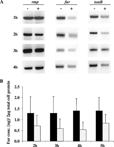
FIG. 1.
In vitro gene and protein expression of gonococcal Fur in iron-replete and iron-depleted growth conditions. (A) Gonococcal gene expression examined by RT-PCR analysis of total RNA isolated from samples collected from iron-depleted (−) and iron-replete (+) conditions, monitored at different time points of growth (indicated to the left). The internal fragments of the iron-regulated fur and tonB genes were amplified and compared to the rmp gene, a gene not regulated by the presence or absence of iron. The amount of RNA utilized for analysis was 150 ng. The amplified cDNA fragments isolated by RT-PCR were run on a 1% agarose gel in 1× TAE buffer with 0.5 μg of ethidium bromide/ml and then visualized under UV light. (B) Gonococcal Fur protein quantification from N. gonorrhoeae F62 grown in iron-depleted (black box) and iron-replete (dotted box) conditions. A total of 2 μg of total cell protein from different growth time points (2 to 5 h) was loaded onto an SDS-PAGE gel and detected by immunoblot analysis using polyclonal anti-Fur antiserum (1:2,000). Fur bands were quantified ± the SD using Adobe Photoshop quantification software (version 6; Adobe Systems Incorporated, San Jose, CA), and the protein concentration was determined by using regression analysis with Fur protein standards of known concentration.

