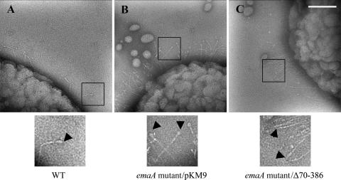
FIG. 3.
Transmission electron micrographs of A. actinomycetemcomitans strains stained with 2% phosphotungstic acid (pH 7). (A) WT, wild type. (B) pKM9, emaA mutant strain complemented with the full-length sequence of emaA. (C) pKMΔ70-386, emaA mutant complemented with a plasmid containing an in-frame deletion corresponding to amino acids 70 to 386. The ellipsoidal endings (arrows) are visualized in both the wild-type strain and the emaA mutant transformed with pKM9, but absent in the emaA mutant complemented with the pKMΔ70-386 construct. The small oval-shaped particles in the micrographs corresponded to membrane vesicles secreted by both wild-type and mutant bacteria. Scale bar, 100 nm.

