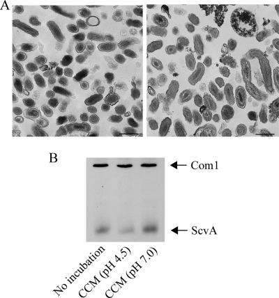
FIG. 8.
Transition of C. burnetii SCV to LCV during incubation in CCM. Purified C. burnetii SCVs were analyzed by transmission electron microscopy before and after a 24-h incubation in CCM to assess potential morphological transitions. (A) SCVs prior to axenic incubation in CCM showed ultrastructural characteristics of this cell form (left panel). Following SCV incubation in CCM, organisms displayed an ultrastructure more characteristic of the LCV form (e.g., size of >0.5 μm and relaxed chromatin; right panel). Bar, 0.5 μm. (B) Scanning densitometry of an immunoblot probed for ScvA (3.5 kDa), a protein specific to SCVs, showed that the ScvA level decreased by approximately 50% following incubation of SCVs in CCM (pH 4.5) for 24 h. SCVs incubated in CCM (pH 7.0) for 24 h exhibited no decrease in ScvA. The levels of Com1 (27 kDa), a protein equally expressed in SCVs and LCVs, did not change. Data from representative experiments are shown.

