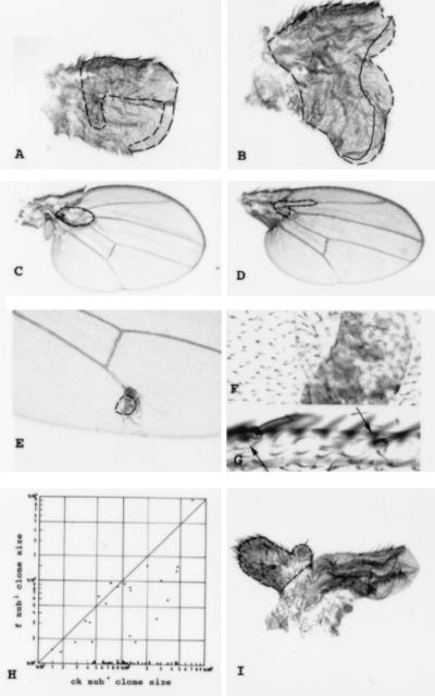
Figure 3.
Cell lineage and morphogenetic mosaics. (A and B) Different examples of early M+ clones (60–108 ± 12 h AEL) which do not respect the A–P and D–V restrictions (dashed line, contour of the dorsal component of the clone; solid line, ventral component). (C and D) nub mutant clones covering the proximal region of the wing blade. Note the small size of the clones (dashed lines, contour of the clone) and the reduction of the whole wing size. (D) Thickening of the proximal L2 is observed. (E and F) Tissue associated with f− cells but located between the two wing surfaces. The trichome pattern of this structure is characteristic of the hinge region (F is a magnification of E). (G) Bracteated bristles (bracts indicated by arrows) in a nub mutant clone covering the triple row. (H) Sizes of nub1 (mwh) clones and of their nub+ (f) twins in double logarithmic representation. (I) nub mutant wing containing a Tubα1>dpp clone (clone border is outlined in dashed lines).

