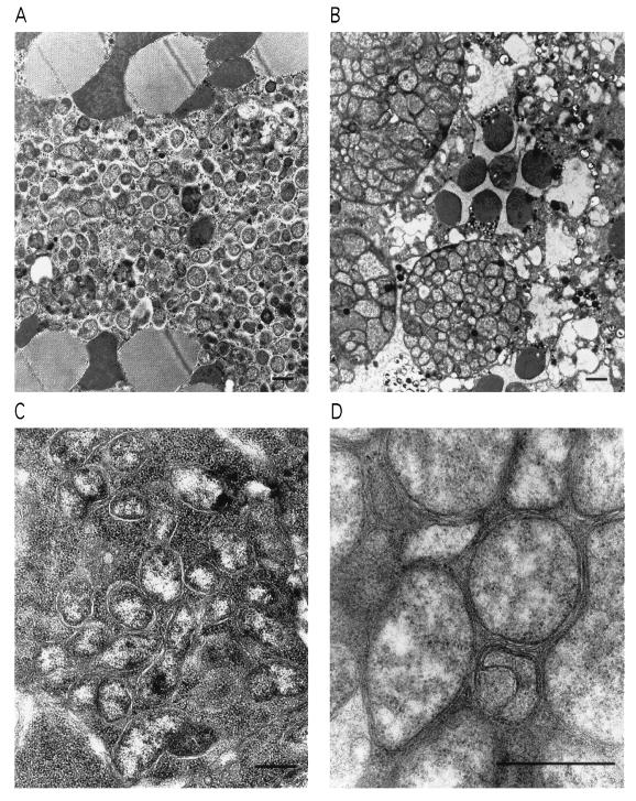
Figure 2.
Proliferation of popcorn in muscle, retina, and ovary. (A) Thoracic indirect flight muscle, seen in oblique section, showing invasion of the bacteria. The large, dark bodies are mitochondria, which deteriorate as the infection progresses. (B) Retina. Masses of the bacteria appear in both photoreceptor cells and surrounding pigment cells. The round, dark bodies are sections of the photoreceptor cell rhabdomeres. (C) Ovarian oocytes. (D) Higher magnification shows the bacterial cell wall and plasma membrane; the morphology is typical of Wolbachia (20). (Bars = 1 μm.)

