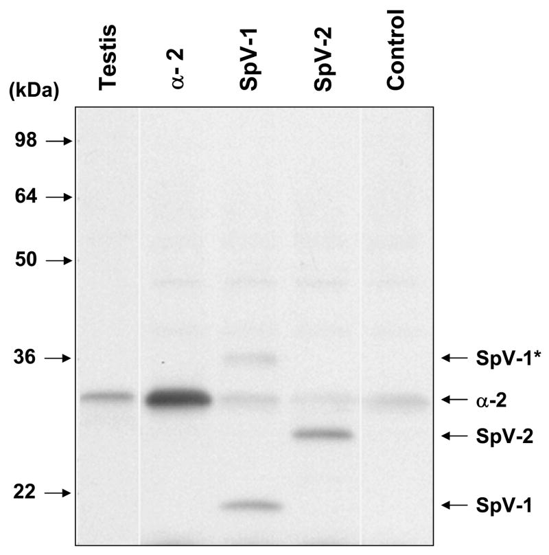
Figure 5.
Pafah1b2 immunostain of in-vitro translated Alpha-2, SpV-1, and SpV-2 separated in a denaturing Tris/Glycine 10–20% acrylamide SDS-PAGE system. In-vitro translations were performed in rabbit reticulocyte lysates using purified cDNA templates produced from human testis RNA. The completed translations were loaded directly on the gel without purifying protein products from the lysate. Chemiluminescent immunostaining is with Pafah1b2 polyclonal primary antibody and a horseradish peroxidase linked secondary. Lane 2 (α2) shows translation from human canonical Pafah1b2 template. Note: endogenous rabbit Pafah1b2 is seen in the control and splice variant translations. SpV-1 and SpV-2 monomers are indicated with labeled arrows. The band corresponding to non-disassociated SpV-1 complex is labeled as SpV-1*. Testis tissue sample (Lane 1) is a total cell lysate of human origin. Control sample is a rabbit reticulocyte lysate transcription/translation reaction performed in the absence of exogenous template.

