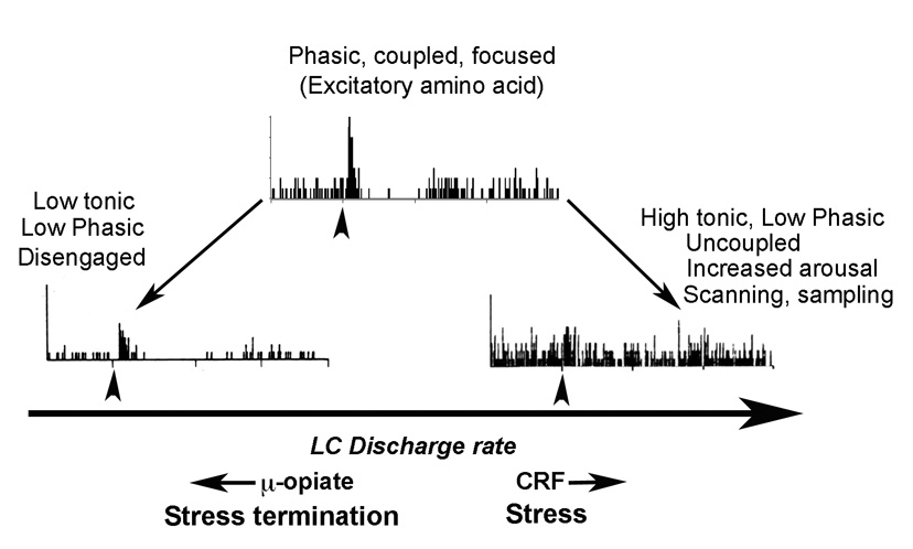
Figure 1.
Schematic depicting different modes of locus coeruleus activity and the relationship between tonic and phasic activity. Representative poststimulus time histograms (PSTHs) are shown that indicate neuronal activation (ordinates) in response to repeated presentations of a brief sensory stimulus (arrowhead). The abscissae of the histograms indicate time before and after the stimulus. When locus coeruleus tonic activity is low (left), the response to sensory stimuli is relatively low and this is associated with drowsiness and disengagement from the environment. There is an optimal level of tonic locus coeruleus discharge rate at which the response to sensory stimuli is high (Phasic). This is associated with electrotonic coupling of locus coeruleus neurons, focused attention and the maintenance of ongoing behavior. Increases in tonic discharge rate above this optimal level result in a loss of selective sensory responses. This is associated with uncoupling, increased arousal, scanning attention and sampling of behaviors. By increasing tonic discharge, CRF shifts the mode of locus coeruleus activity towards the right of this spectrum. This is predicted to occur during stress. During stress termination, endogenous opioids acting at μ–opiate receptors in the locus coeruleus shift the mode of activity towards the left. Excitatory amino acid neurotransmission in the locus coeruleus may favor the phasic mode.

