
Figure 5.
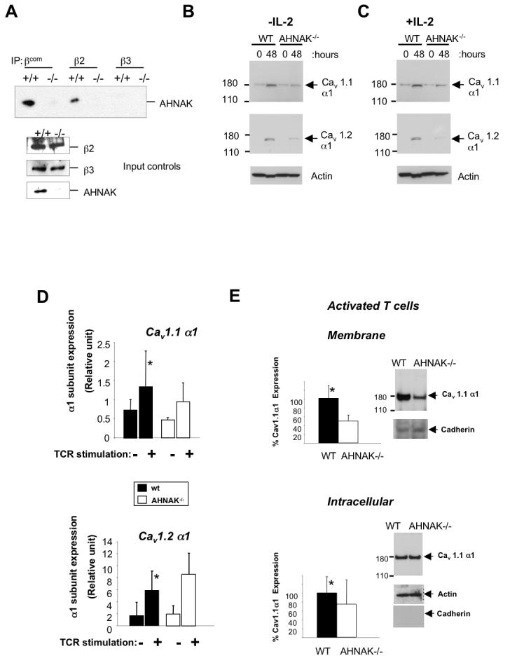
AHNAK1 is required for membrane expression of Cav1 channels. (A) Association of AHNAK1 with the β subunits of Cav1 channels. Lysates were prepared from splenocytes of wild-type and AHNAK1−/− mice and immunoprecipitated with antibodies against β subunit 2, 3 or all βsubunits (βcom). The membrane was blotted with anti-AHNAK1-C2 antibody. Results are representative of two independent experiments. (B) CD4 T cells were stimulated with anti-CD3 (10 μg/ml) and anti-CD28 (2 μg/ml) antibodies and total cell lysates were prepared. The expression of Cav1 channels was examined by western blotting with anti-Cav1.1 and Cav1.2 antibodies. The same membrane was blotted with anti-β-actin antibody for loading control. Results are representative of at least three independent experiments. (C) This experiment was performed as in B with the only difference that IL-2 was added to the culture media. Results are representative of at least three independent experiments. (D) CD4 T Cells were stimulated as in C and Cav1.1 and 1.2 α1 subunits expression was examined by real time PCR. Results are representative of at least three independent experiments. p-value represent the difference in expression between wild-type and AHNAK1−/−(p=0.235 and p=0.366 for Cav1.1 and Cav1.2, respectively). (E) Cells were stimulated as in C followed by fractionation of membrane proteins and western blot analysis as described in B. Densitometry represents statistical analysis of three independent experiments (p=0.02 and p=0.25 for membrane and intracellular expression of Cav1.1 respectively). Cav1.1 membrane protein expression was normalized to pan-cadherin and intracellular expression was normalized to β-actin. A representative western blot used for densitometry is shown.