
Figure 3.
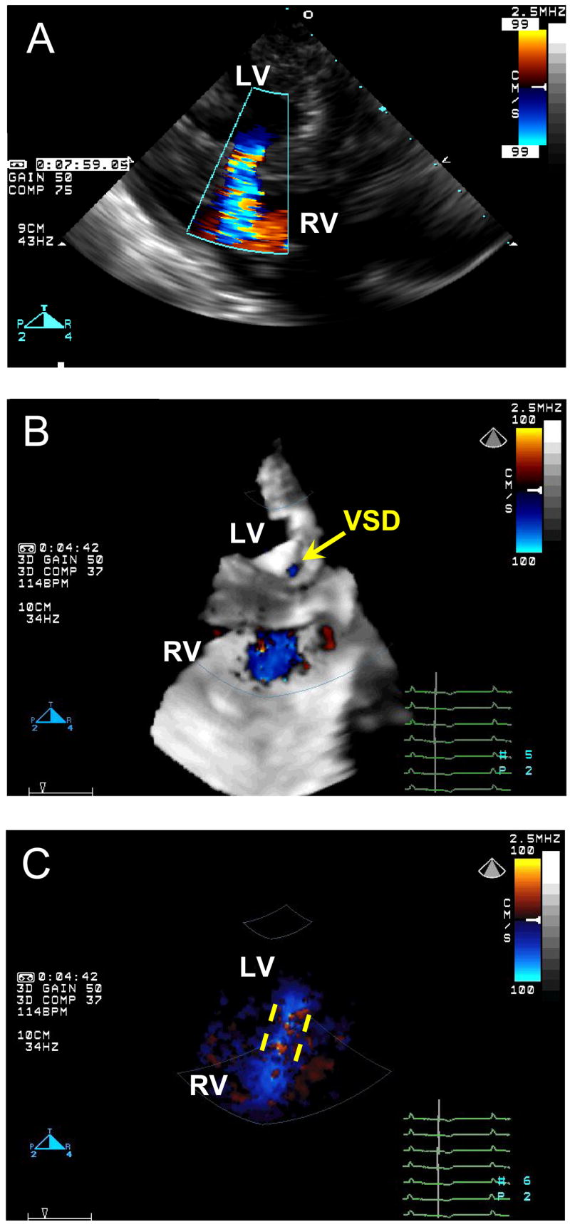
Color Doppler images of the created VSD. A, 2D Color Doppler. B, 3D Color Doppler, yellow arrow points to the orifice of the VSD on the left side of the ventricular septum. C, the same 3D Color Doppler image with the black-and-white suppress, yellow dashed lines demonstrate the VSD tunnel inside the ventricular septum. LV, left ventricle; RV, right ventricle; VSD, ventricular septal defect.