
Figure 2.
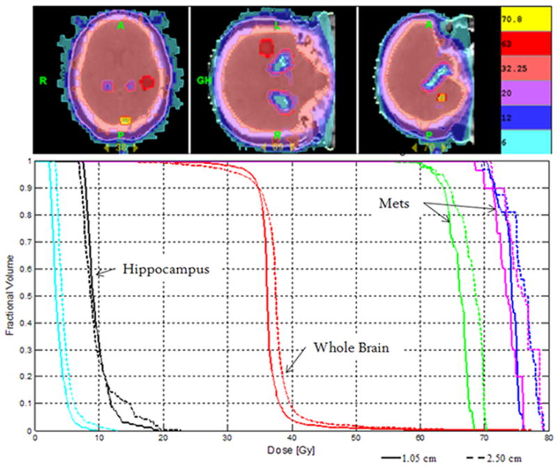
(Top) Example isodose distribution of WBRT-HA with SIB of three metastases using helical tomotherapy. (Bottom) Corresponding cumulative, normalized DVH for WBRT-HA with SIB to three metastases. Two metastases prescribed to 70.8 Gy (magenta & blue), one metastasis to 63 Gy (green), whole brain (red) to 32.25 Gy. Hippocampus (black) and eyes (cyan) are shown. Dashed line represents plan with 1.0cm FW, and solid line represents plan with 2.5cm FW—both pitch of 0.289. Plans normalized to prescription dose for comparison purposes.