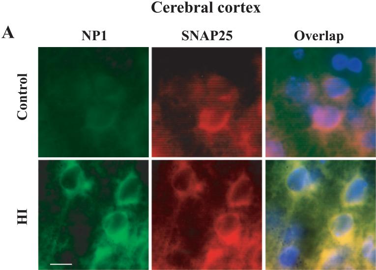
Figure 3.
NP1 induction in neonatal brain following HI is neuronal specific. Brain sections (15 μm) collected from sham controls and HI animals sacrificed at 24 h post-HI. Immunofluorescence staining was performed with respective primary antibody for NP1 (1:500), SNAP25 (1:500) or GFAP (1:1000) followed by incubation with appropriate FITC- (green) and Texas red- (red) conjugated secondary antibodies. Brain sections were coverslipped with Prolong Gold (Invitrogen) antifed mounting medium containing 4,6diamino-2-phenylindole (DAPI) that stains nuclei (blue). Immunofluorescence analyses were performed using a fluorescence microscope (Carl Zeiss Axioplan 1 microscope fitted with AxioVision 3.0 software). Representative images are shown taken from cerebral cortex ischemic penumbra and viewed at 100 × magnifications. Bar = 10 μm.

