
Figure 1.
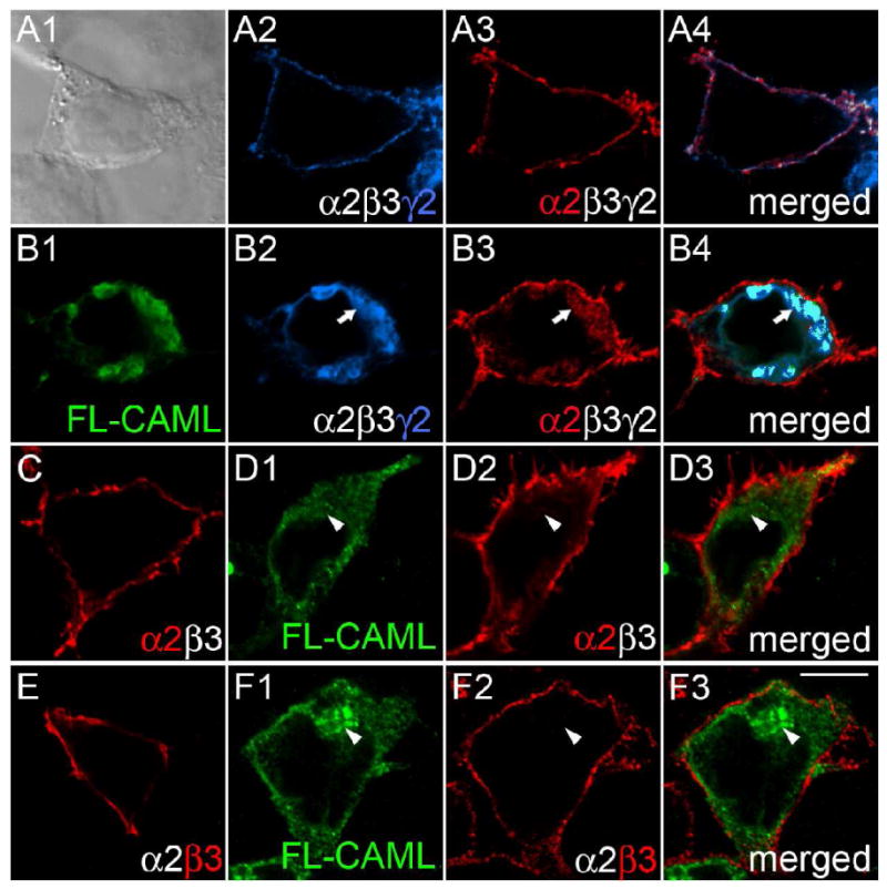
CAML interacts with the γ2 but not α2 and β3 subunits in transfected 293T cells. A-F, Human embryo kidney 293T cells were transfected with α2β3mycγ2 (A, B) or α2β3 receptors (C - F), either alone (A, C, E) or together with FL-CAML (B, D, F) and stained with antibodies specific for the mycγ2 subunit (A2, B2, blue), the α2 subunit (A3, B3, C, D2, red) or β3 subunit (E, F2) and FL-CAML (B1, D1, F1, green). Panel A1 shows a bright field image; the other panels show single optical sections of representative cells imaged by confocal microscopy. Merged images representing two or three separate fluorescent channels of cells in A, B, D, and F are shown in the right column. Note the colocalization of α2 and γ2 subunits at the cell surface of the α2β3mycγ2 receptor-transfected cell in (A) evident in the merged image (A4) that is absent upon cotransfection of CAML with α2β3mycγ2 receptors in the cell shown in (B) (merged image B4). Whereas cells cotransfected with FL-CAML and α2β3mycγ2 show intracellular trapping of the γ2 (B2, B4) and α2 subunits (B3), virtually no intracellular GABAAR subunit staining is seen when α2β3mycγ2 receptors are transfected alone (A2-4), or upon cotransfection of α2β3 receptors with FL-CAML (D2, D3, F2, F3). Arrows point to colocalization of FL-CAML and GABAAR subunits, arrowheads indicate lack of colocalization. Scale bar, 10 μm.