
Figure 4.
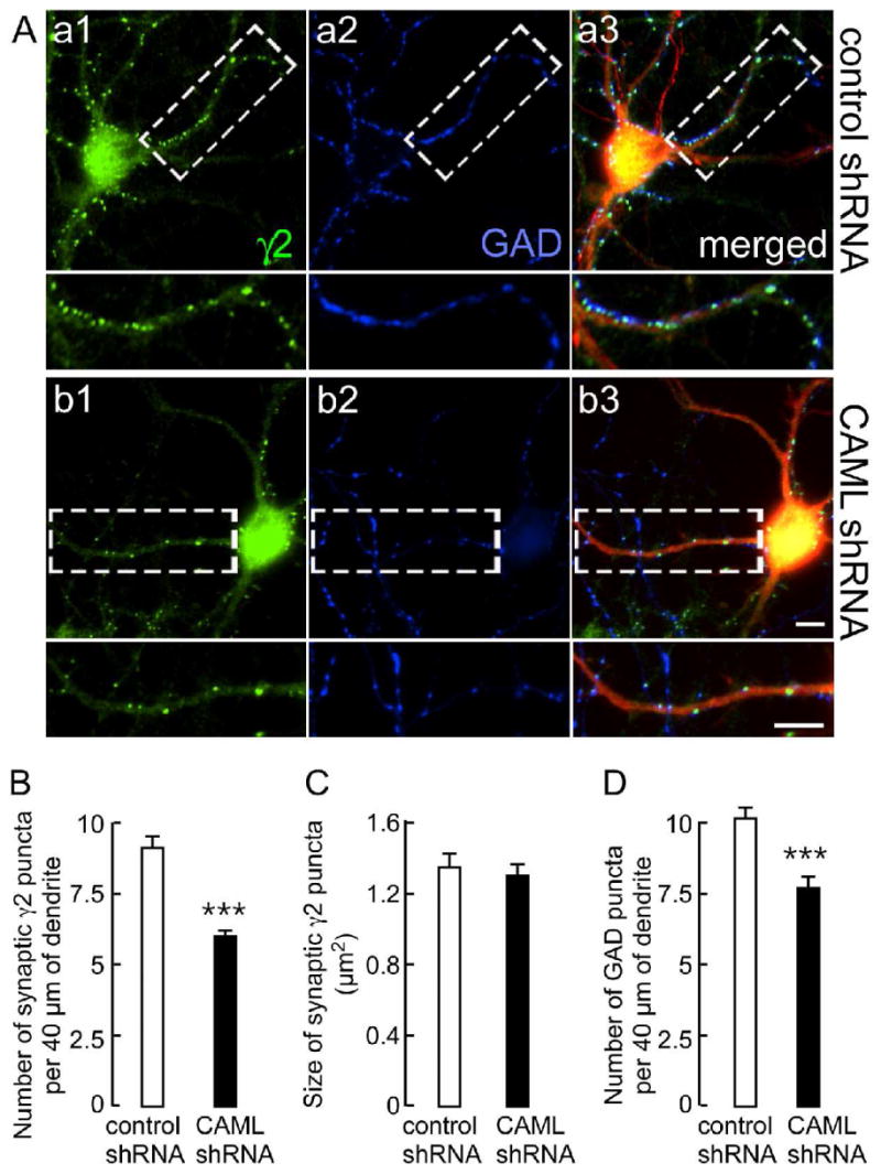
CAML shRNA interferes with expression of postsynaptic γ2 subunit-containing GABAARs and, indirectly, with GABAergic innervation. A. Cortical neurons transfected with control shRNA (Aa1-Aa3) or CAML shRNA (Ab1–Ab3) were stained for the γ2 subunit (green, Aa1, Ab1) and GAD (blue, Aa2, Ab2). Transfected dendrites were identified based on expression of DsRed encoded by the shRNA plasmids, as evident in the merged images (red). Boxed dendritic segments (40 μm) are shown enlarged in the panels below for either γ2, GAD alone, or as merged images. Scale bars, 5 μm. B-D. Semiquantitative analyses of the number (B) and size (C) of immunoreactive puncta for the γ2 subunit apposed to GAD and the number of GABAergic terminals immunopositive for GAD (D) in 40 μm of dendritic segments. Note the prominent reduction in the number of γ2 subunit puncta (CAML shRNA: 6.0 ± 0.2, n = 110; control shRNA: 9.1 ± 0.4, n = 47 cells, p < 0.001) but unaltered size (CAML shRNA: 1.30 ± 0.07 μm2, n = 109; control, 1.35 ± 0.08 μm2, n = 47, p = 0.41). Similarly, the number of GAD puncta was significantly reduced (CAML shRNA, 7.7 ± 0.4; control, 10.1 ± 0.4, n = 25, p < 0.001). Data represent means ± S.E.; ***, p < 0.001, Student t-test.