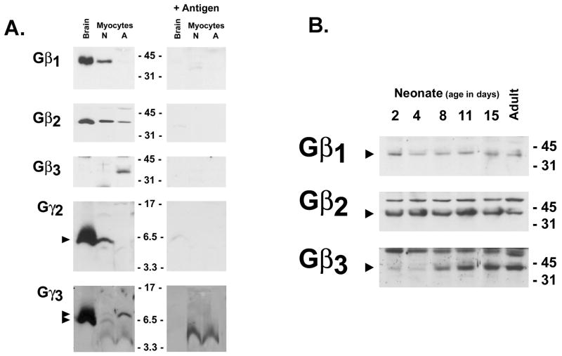
Figure 1. Developmental changes in Gβ and Gγ expression in the ventricle.
100 μg of total cell protein extracted from day 5 neonatal cardiomyocyte cultures, or freshly isolated adult rat cardiomyocytes (Panel A) or from ventricles from rats at the indicated ages (Panel B) or were subjected to SDS-PAGE and immunoblot analysis with the indicated antibodies. An extract from brain (100 μg) was included as a positive control in Panel A. Epitope specific immunoreactivity was established by immunoblotting with antibody complexed with competing antigen peptide; note smaller proteins detected by the Gγ3 antibody are non-specific. Arrow denote epitope specific bands, with positions of the molecular weight standards (in kDa) indicated.

