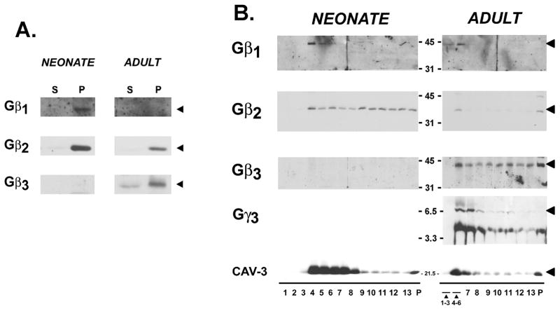
Figure 2. Gβ and Gγ partitioning to membranes and low-density caveolae in cardiomyocytes.
Panel A: Neonatal cardiomyocyte cultures and acutely isolated adult cardiomyocytes were partitioned into soluble and particulate fractions and then subjected to immunoblot analysis for Gβ subunits as indicated. Panel B: Neonatal cardiomyocyte cultures and isolated adult cardiomyocytes were homogenized in sodium carbonate buffer and subjected to sucrose gradient centrifugation as described in methods. Fractions were collected from the top of the gradient and analyzed by SDS-PAGE and immunoblot analysis with the indicated antibodies. Fractions 1–3 and 4–6 in profiles from adult cardiomyocytes were pooled, due to the limiting amounts of protein recovered in these fractions. Immunoblot analysis was on 35 μg of protein from each fraction. Results are representative of data from three separate experiments.

