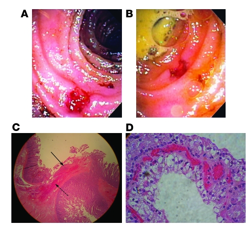
Figure 2. Gross and histologic pathology.
(A and B) Intraoperative endoscopy shows a well-demarcated ulcer in the proximal ileum with minimal surrounding inflammation. (C) A shallow jejunal ulcer involves the mucosa and submucosa. Acute inflammatory exudate covers the ulcer base (solid arrow). Neutrophils infiltrate the submucosa (dashed arrow). There are no signs of chronicity nor evidence of viral inclusion, inflammatory bowel disease, or vasculitis. Original magnification, ×100. (D) Renal-cell carcinoma, Furman grade II. Original magnification, ×100.

