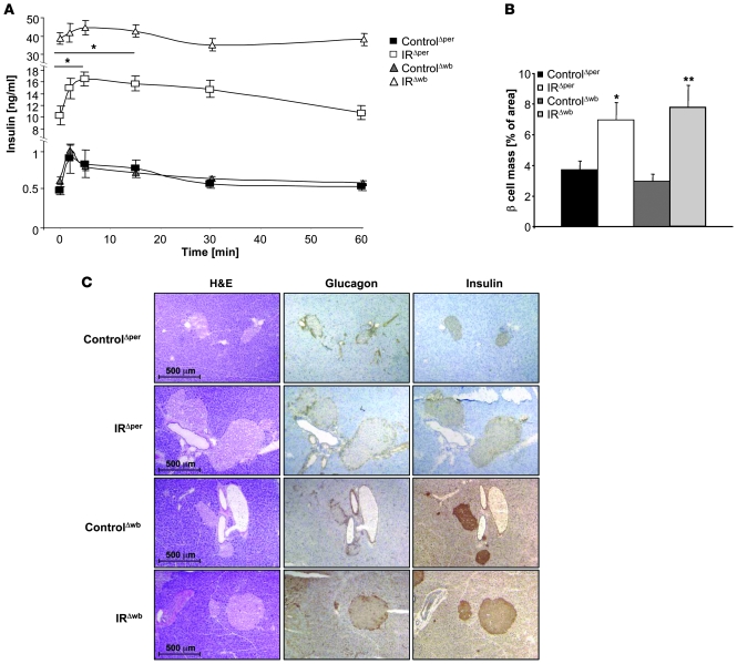
Figure 9. Plasma insulin levels and glucose-stimulated insulin secretion in IRΔper and IRΔwb mice.
(A) Glucose-stimulated insulin secretion of 13-week-old ControlΔper (filled squares; n = 8), IRΔper (open squares; n = 5), ControlΔwb (filled triangles; n = 5), and IRΔwb mice (open triangles; n = 8) on day 18. Values are mean ± SEM. *P ≤ 0.05 versus 0 min. (B) Percentage of β cell mass in 14-week-old ControlΔper (black bars; n = 4), IRΔper mice (white bars; n = 4), ControlΔwb (dark gray bars; n = 3), and IRΔwb mice (light gray bars; n = 3). Data was collected 30 days after starting the experiment. Values are mean ± SEM. *P ≤ 0.05, **P ≤ 0.01 versus control. (C) Immunohistochemical stainings of pancreatic islets in 14-week-old ControlΔper, IRΔper, ControlΔwb, and IRΔwb mice. Pancreatic tissues were stained for H&E, insulin, and glucagon. Magnification, ×100.

