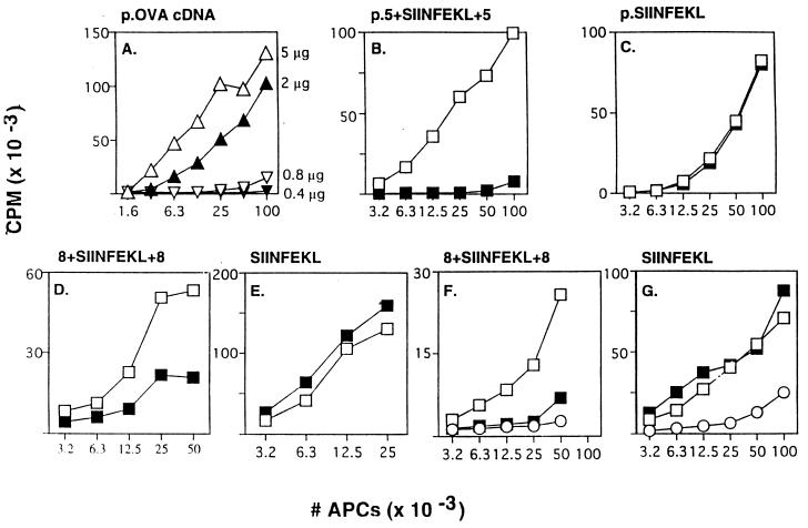
Figure 1.
MHC class I antigen presentation from OVA protein and SIINFEKL-containing oligopeptides. (A) E36.12.4 APCs were infected with vTF7–3 and then transfected with the indicated concentrations of plasmid encoding intact OVA cDNA. Assays for presentation of SIINFEKL on Kb were performed as described (7). (B) Similar to A, except that cells were treated with (▪) or without (□) lactacystin (2 μM) for 30′ before transfection and during the subsequent incubation, and 5 μg of a plasmid encoding p.5+SIINFEKL+5 was transfected. (C) Same as B except that the plasmid encoded p.SIINFEKL (0.7 μg transfected). (D) Similar to B except that 8+SIINFEKL+8 synthetic peptide (300 μg/ml) was introduced into the cytosol of LB27.4 cells (17) (APCs) by electroporation (7) (instead of vaccinia infection and plasmid transfection) and lactacystin was used at 40 μM. (E) Same as D except that SIINFEKL (0.5 μg/ml) was used instead of 8+SIINFEKL+8 peptide. (F) Same as D except that LLnL (40 μM) was used instead of lactacystin. (G) Same as E except LLnL (40 μM) was used instead of lactacystin. Some APCs were fixed immediately after electroporation of antigen (○) to rule out peptide binding directly to cell surface MHC molecules (7). All results in Figs. 1, 2, 3, 4 are representative of data obtained in repeated experiments. OVA constructs expressed from plasmids are indicated with the prefix p.

