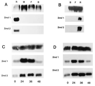
Figure 4.
Northern analysis. Each of the membranes (A-D) were probed once with Smd1, stripped, and reprobed with Smd2. (A) Specific tissues from flies fed on pig blood containing 3,000 units/ml LPS. (Upper) Ethidium bromide staining of RNA. (B) Specific tissues from flies injected with LPS. (Upper) Methylene blue staining of RNA. (C) Whole fly extracts at the indicated hours after feeding on sterile, heparinized pig blood. (Upper) Aedes aegypti ribosomal probe. (D) Whole fly extracts at the indicated hours after feeding on heparinized pig blood plus LPS at 3,000 units/ml. (Upper) A. aegypti ribosomal probe. R, anterior midgut; M, posterior midgut; F, fat body; B, remainder of the carcass.

