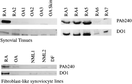
Figure 2.
p53 protein detection by immunoprecipitation. p53 protein expression was analyzed in synovial tissue (RA and OA), skin (OA), fibroblast-like synoviocytes [RA, OA, and normal (NML)], and dermal fibroblasts (DF). Different samples are from different patients and are numbered arbitrarily. Immunoprecipitation was performed as described in Methods using either PAb240 (detects mutant but not wild-type p53) or DO1 (detects mutant and wild-type p53). Note the discordant expression of DO1 and PAb240 precipitable protein in OA joint samples, whereas abundant p53 was detected in RA joint tissue using PAb240.

