
Figure 1.
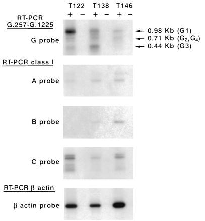
Detection of HLA class I transcripts in cytotrophoblast cells isolated in vitro from first trimester terminations of pregnancy. Reverse RNAs were amplified using either G.257 and G.1225 HLA-G-specific primers or pan class I-specific primers, and Southern blots were hybridized with either an HLA-G 32P-labeled probe (G probe), an HLA-A 32P-labeled probe (A probe), an HLA-B 32P-labeled probe (B probe), or an HLA-C 32P-labeled probe (C probe). Positive (+) and negative (−) lanes correspond to the RT+ and RT− template, as described. To control the amount of RNA in the samples, RT-PCR amplification results obtained with β-actin-specific primers and Southern blots were hybridized with a β-actin 32P-labeled probe. Note that all the cytotrophoblasts present HLA-G1, -G2, -G3, and -G4 transcripts, but at various levels.