Figure 4.
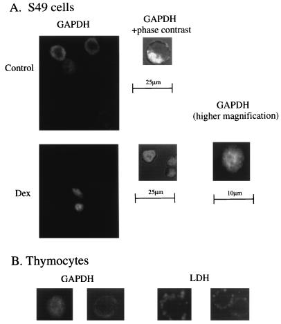
Translocation of GAPDH to the nucleus; immunocytochemical studies in S49 cells and primary thymocytes. (A) Prior to Dex treatment, GAPDH staining is excluded from the nucleus in S49 cells. (Left) A confocal image of GAPDH staining. (Right) A confocal section of the phase contrast image superimposed on GAPDH staining. Nuclear exclusion is detected in almost all of more than 500 cells observed. After Dex treatment, GADPH staining is uniform over S49 cells. These pictures were taken 48 hr after stimulation, when more than 70% of cells are still intact by trypan blue exclusion assay. Left shows a confocal image of GAPDH staining. Center shows a confocal section of the phase contrast image superimposed on GAPDH staining. Right shows GAPDH staining at higher magnification. No nuclear exclusion of GAPDH is detected in more than 500 cells examined with independent confocal sections at 0.5-μm intervals. (B) In primary thymocytes treated with Dex, GAPDH staining is uniformly distributed in nuclei and cytosol, whereas antisense oligonucleotide treatment (5 μM) restores nuclear exclusion (Left). LDH staining is nonnuclear both before and after Dex treatment. After stimulation, cell size is reduced slightly with no change in the staining pattern of LDH (Right).

