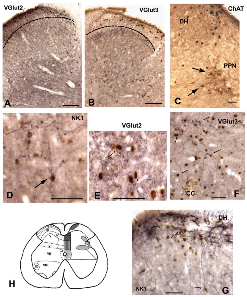
Figure 3.
Photomicrographs illustrating the colocalization of fos-I nuclei with VGlut2 (A, E), VGlut3 (B, F), NKI (D, G) and ChAT (C). [A and B] Show the dorsal horn, the dotted line represents the approximate border of lamina II and lamina III. VGlut2 immunoreactivity is more prominent in the superficial laminae where it is co-distributed with fos-I nuclei. VGlut3 immunoreactivity is predominate in laminae III and not associated with fos-I nuclei in the superficial dorsal horn. [C] ChAT and fos-I nuclei in the dorsal horn and lateral gray. Arrows shows examples of cells colocalized with c-fos and ChAT in the PPN. [D and G] Examples of NKI activated cells in the dorsal horn (arrows). [E] VGlut2 immunoreactive dendrites and boutons adjacent to fos-I nuclei in the lateral gray (arrows). [F] VGlut3 immunoreactive dendrites and boutons associated with fos-I nuclei in the dorsal horn (arrows). Abbreviations:- DH - dorsal horn, CC -central canal, PPN - parasympathetic preganglionic nucleus. Scale bars = 250μm for A and B; 100μm for C-G. [H] Illustrates a summary diagram showing areas that resulted in increased fos-immunoreactivity after both pudendal sensory and pelvic nerve stimulation. The relationship of these areas with NKI, VGlut2 and VGlut3 is also shown. Left side shows the spinal laminae. Right side shows shaded areas in which fos-immunoreactivity was significantly increased with both pudendal and pelvic nerve stimulation. Specific shading subcategorizes areas showing significant overlap of fos-I nuclei with NKI, VGlut2 or VGlut3. Dark grey = NKI+VGlut2; vertical lines = VGlut3; diagonal lines = NKI; horizontal lines = VGlut2; and light shading = neither NKI, VGlut2 nor VGlut3.

