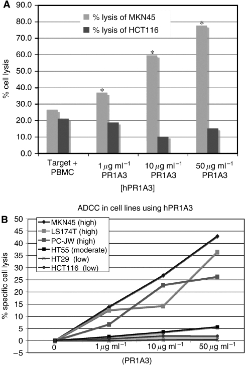
Figure 4.
hPR1A3-mediated ADCC lysis of colorectal cancer cell lines depends on their level of CEA expression. (A) Effect of increasing concentrations of hPR1A3 on lysis of CEA-positive (MKN45) and -negative (HCT-116) cell lines. Fluorescence-based ADCC assays were done using human PBMCs as effector cells in a ratio of 100 : 1 with target cells. Columns represent % lysis in the presence of both target and effector cells with no, or with increasing concentrations of hPR1A3. *P<0.05 comparing the cell lysis between MKN45 and HCT116. (B) Comparison of hPR1A3-mediated ADCC based lysis in cell lines with different levels of CEA expression (shown in parentheses) based on results in Table 1. The EuTDA-based ADCC assay was done using human PBMCs as effector cells at ratios of 100 : 1 with the various target cell lines. Nonspecific spontaneous killing levels have been subtracted to reflect antibody-specific lysis only.

