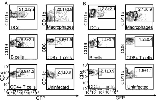
Figure 1.
MVA preferentially targeted professional APCs, especially DCs, both in vitro and in vivo. (A) Splenocytes from naïve BALB/c mice were infected with rMVA-GFP at a MOI of 10 in vitro for 8 h followed by cell surface marker staining and flow cytometry analysis. Uninfected splenocytes were used as negative control. (B) BALB/c mice were either uninfected or infected with rMVA-GFP at 3 × 10⁸ PFU/mouse by i.v. injection. Spleens were harvested at 9 h post infection and GFP expression in various subsets of splenocytes was monitored by flow cytometry. DCs: CD11c+; macrophages: CD1b+CD11c-; B cells: CD19+; CD8+ T cells: CD8+ CD11c-; CD4+ T cells: CD4+ CD11c-. Numbers shown are the percentages (average ± standard deviation (SD)) of GFP+ cells in the corresponding cell subsets. The data are representative of three independent experiments.

