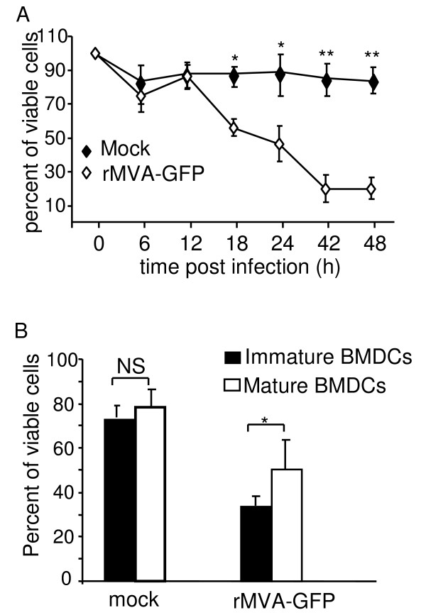
Figure 5.
MVA infection significantly reduced the viability of BMDCs after 12 h of infection. (A) Kinetic analysis of DC viability following MVA infection. Immature BMDCs were either mock treated, or infected with rMVA-GFP at a MOI of 10. At various time points, cells were harvested and viable cells were counted with trypan blue exclusion method. These data represent the average ± SD viability of 6 replicates from 2 experiments. (B) Immature BMDCs and LPS-stimulated mature BMDCs were mock treated or infected with rMVA-GFP at a MOI of 10. Twenty four hours later, cell viability was examined by trypan blue exclusion. These data represent the average (± SD) viability of 15 replicates from 5 experiments. NS: statistically non-significant; *: P < 0.05; **: P < 0.01.

