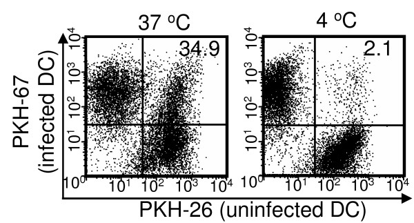
Figure 6.
MVA-infected DCs were phagocytosed by uninfected DCs. Immature BMDCs were labeled with PKH-67 and infected with MVA at a MOI of 10 for 18 h. They were then extensively washed and mixed with PKH-26-labeled uninfected immature BMDCs for 4 h at either 37 C° or 4 C°. Phagocytosis of MVA-infected DCs by uninfected DCs were detected by flow cytometry. This data is representative of two independent experiments.

