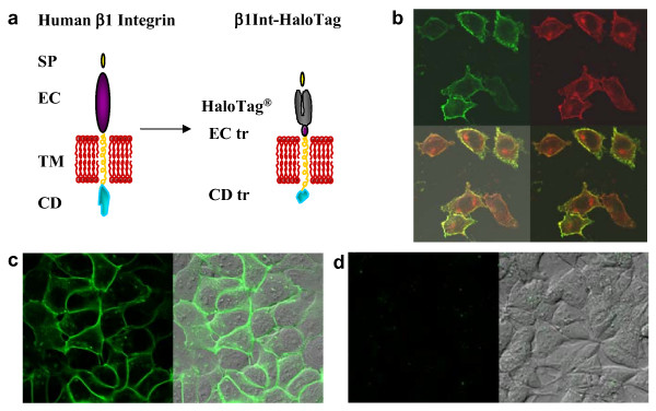
Figure 2.
Targeting HaloTag protein to the cell surface. (a) Human β1 integrin is depicted with a signal peptide (SP), extracellular domain (EC), transmembrane domain (TM) and cytoplasmic domain (CD) (left). In the β1Int-HaloTag construct, the HaloTag protein is displayed on the cell surface by fusion to the truncated (tr) extracellular domain of human β1 integrin (right). (b) Immunocytochemistry, without permeabilization, using antibodies against integrin (green) and HaloTag (red) show that the β1Int-HaloTag fusion protein is being expressed in transfected HeLa Hcells, and the overlap of the two proteins (yellow) indicates β1Int-HaloTag is expressed on the cell surface in a similar pattern to endogenous β1 integrin. (c) HEK293 cells stably expressing β1Int-HaloTag labeled with the HaloTag 488 ligand show a green rim, confirming that HaloTag is on the surface and functional. (d) HEK293 cells stably expressing HaloTag-(NLS)3 labeled with the HaloTag 488 ligand do not show a green rim, confirming that the ligand is cell impermeable and does not label intracellular HaloTag protein. Cell images were generated on an Olympus FV500 confocal microscope using the appropriate filter.

