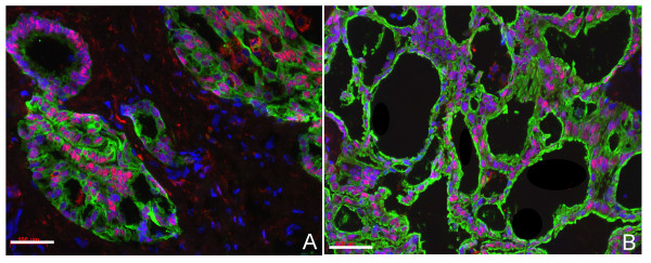
Figure 3.
Double immunostaining of Prox1 (red) with OV-6 or CK19 (green) in intrahepatic cholangiocellular carcinoma. (A) OV-6; (B) CK19 show duct-like glandular or acinar structures localised within a dense fibrous stroma. Note that the cells are positive for OV-6 or CK19 and for Prox1. The blue colour staining with DAPI represents the nuclei. Bars represent 100 μm.

