
Figure 4.
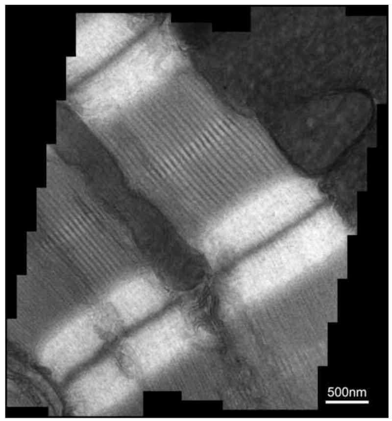
The mosaic image (~2k x 2k) of longitudinal mouse cardiac muscle specimen from 44 individual 512 x 550 pixel DDD images, each taken at 2500 magnification. For each image, the total dose is <200 electrons/pixel. The main features of the cardiac muscle, including the muscle Z line, myosin bundles and mitochondria, are clearly visible.