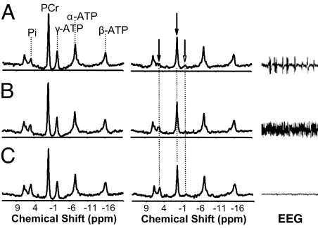
Fig. 1.
In vivo 31P MT and EEG measurements from a representative rat brain under IsoF (SEI = 0.75) (A), low-Pen (SEI = 0.65) (B), and high-Pen (isoelectric; SEI = 0.50) (C) anesthesia conditions, respectively. (Left and Center) 31P spectra acquired in the absence (control) (Left) and presence (Center) of γ-ATP saturation. The magnetization ratio quantified by the NMR signals obtained at steady-state saturation versus control was 56%, 59%, and 63% for PCr and 59%, 67%, and 78% for Pi at IsoF, low-Pen, and high-Pen anesthesia conditions, respectively. The ratio changes indicate reduction in the measured ATP metabolic rates with increased anesthesia depth. (Right) EEG time courses recorded at three anesthesia conditions.

