Fig. 5.
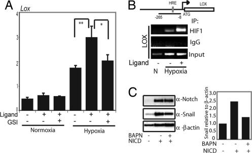
Notch signaling enhances hypoxia-induced activation of LOX transcription. (A) Quantitative PCR analysis of LOX expression in SKOV-3 cells cocultured with 3T3-Babe (3T3-B) or 3T3-Jagged (3T3-J) cells kept at normoxia or treated with hypoxia for 16 h in the absence or presence of GSI. Values are significant at **, P < 0.01 and *, P < 0.05 as indicated. Values represent the average of three independent experiments. (B) Schematic depiction of the LOX promoter with the PCR-amplified promoter region and the location of the HRE sites denoted. For the ChIP experiments, SKOV-3 cells were cocultured with either 3T3-J cells or 3T3-B cells at normoxia or hypoxia, as indicated. PCR amplification of the LOX promoter after immunoprecipitation of HIF-1α. (C) Western blot analysis of Snail-1 expressed from a heterologous promoter to analyze Snail-1 protein stability. SKOV-3 cells were transfected with wild-type Snail-1 and N1ICD or an empty vector and subjected to hypoxia in the presence or absence of the LOX inhibitor BAPN (Left). Quantification of the Western blot (Snail-1/β-actin intensity) is shown (Right).

