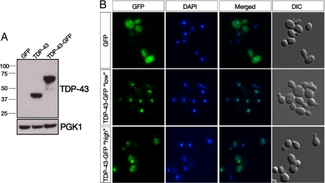
Fig. 1.
Yeast TDP-43 proteinopathy model. (A) TDP-43 or TDP-43-GFP expression in yeast cells was detected by immunoblotting with a mouse polyclonal TDP-43 antibody. Phosphoglycerate kinase 1 (PGK1) was used as a loading control. (B) Fluorescence microscopy was used to visualize the subcellular localization of C-terminally GFP-tagged TDP-43 fusion proteins. Cells were stained with DAPI to visualize nuclei. Whereas GFP alone was distributed between the cytoplasm and nucleus (Top), one integrated copy of TDP-43-GFP strongly localized to the nucleus (Middle) with the occasional formation of intranuclear foci. Expressing TDP-43-GFP from a 2-μm (2 μ) high-copy plasmid profoundly altered its localization because the majority of TDP-43 was now found in multiple cytoplasmic foci (Bottom).

