Fig. 1.
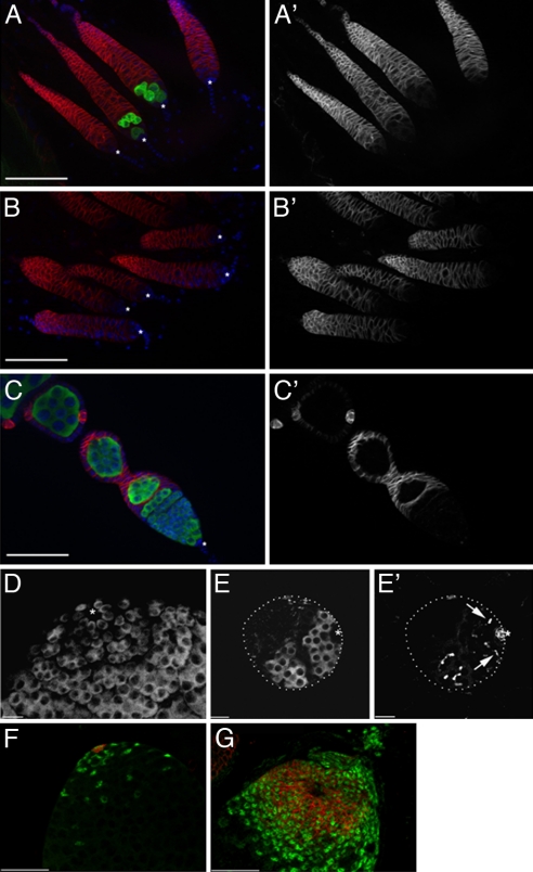
GC loss and expansion of somatic cells in gonads from flies misexpressing bam+. (A–C) GC loss in adult females misexpressing bam+ in the germ line (y w/w1118;UASp-bam+::gfp/+; nos-GAL4::VP16/+). GCs are stained for GC specific antigen vasa (green), somatic cells (FasIII, red), and DNA (DAPI, blue). Asterisk (*) denotes somatic cap cells. (A) GCs are rarely observed in ovarioles from 2-day-old females overexpressing bam+ [24/225 ovarioles (10.7%) contained GCs]. (B) GC loss is virtually complete by 7 days [3/325 ovarioles (0.9%) contained GCs]. See also Fig. S1. (C) Germarium from a 2-day-old control female. All ovarioles from control females contained GCs at 2 (n = 120) and at 7 days (n = 152). (A′–C′) shows FasIII+ somatic cells only. Note the expanded somatic gonad in GC-less females. (D and E) GC loss in third instar larval (L3) males overexpressing bam+. Control testes from L3 males (D) have a normal number of GSCs (vasa) in contact with hub cells (*) at the apical tip. (E) Males overexpressing bam+ show loss of GSCs by this stage. (E′) GCs present near the hub have branched fusomes (arrows) as detected by staining with antibodies to α-spectrin. Note the reduced size of the gonad. Asterisk (*) denotes the FasIII+ apical hub. (F and G) Expansion of somatic cells in testes from adults misexpressing bam+. (F) Control testes show normal distribution of FasIII+ hub cells (red) and TJ+ somatic cells (green). (G) An expansion of FasIII+ and TJ+ somatic cells is observed in testes. (Scale bars: 50 μm in A–C, F, and G; 20 μm in D and E.)

