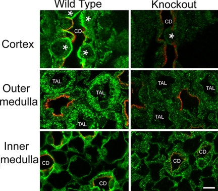
Fig. 4.
Immunofluorescent localization of IR-β. Dual labeling of IR-β (green) and aquaporin-2 (AQP2, red), marker for collecting duct principal cells in the three regions of the kidney in WT and KO mice. IR immunofluorescence was markedly diminished in the cortex (Top) collecting duct (CD) cells, both principal cells (labeled red with AQP2 antibody) and intercalated cells (indicated with an asterisk). In the outer medulla (Middle), IR immunofluorescence was also diminished in the thick ascending limb cells (TAL). In the inner medulla (Bottom), IR was strongly reduced in the inner medullary collecting duct cells (CD). (Total magnification, ×1,000.)

