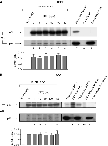
Figure 2.
Steroid receptors AR and ERα interact with p85/PI3K in LNCaP and PC-3 prostate cancer cells and such interaction is not altered by RES treatment. LNCaP (A) and PC-3 (B), growing in complete medium, were treated with the indicated concentrations of RES for 36 h and 1 mg total cell extracts immunoprecipitated (IP) with anti-AR- or anti-ERα-specific antibodies, respectively. The amount of p85/PI3K associated to each receptor was determined in the immunoprecipitates by Western immunobloting (WB) (lower blots in A and B, lanes 1–6). For quantitation, immunoprecipitated p85 was normalised by the amount of AR (A) or ERα (B) at each concentration of RES in three different cultures (lanes 1–6). For LNCaP cells (A) total cell extract was used as a positive control for AR and p85 expression (lane 7), whereas negative controls for this cell line included AR immunoprecipitation (lane 8) and total extract (lane 9) from androgen-insensitive PC-3 cells. For PC-3 (B), positive controls for ERα and p85 expression included total cell extracts from this cell line (lane 7) and from human breast tumour MCF-7 cells (lane 9); for ERα association to p85, immunoprecipitations were carried out in oestrogen-responsive MCF-7 (lower blot in B, lane 8). Negative controls were also included for ERα expression (lane 11) and for its interaction with p85 (lane 10) using oestrogen-unresponsive human breast tumour MDA-MB-231 cells. As an additional negative control, immunoprecipitations were carried out in the absence of AR or ERα antibodies (no antibody in A and B).

