Figure 3.
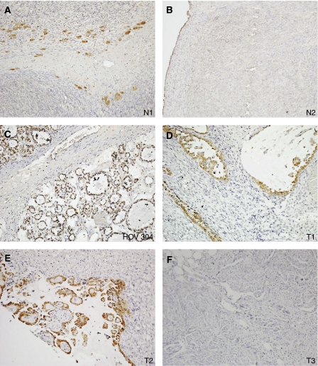
Immunohistochemistry of normal ovary (A, B) and ovarian cancer (C–F) sections. Staining in the normal ovary can be seen to be confined to luteinised stromal cells (N1) and low level expression in the ovarian surface epithelium (N2). HOV304 corresponds to one of the clear-cell tumors that was also analysed by RT–PCR and T1 and T2 are clear-cell tumours from the histoarray. T3 is a negative staining tumour. Magnification × 10.

