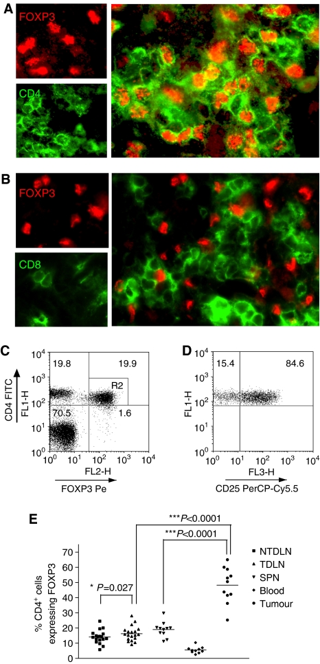
Figure 1.
Methylcholanthrene-induced tumours exhibit a significant enrichment of CD4+FOXP3+ Tregs. Sections of MCA-induced tumours were stained with anti-FOXP3-specific Ab and either anti-CD4 (A) or –CD8 (B) specific mAb. Single-cell suspensions of TILs, NTDLN, TDLN, spleen and blood were stained with anti-CD4-, anti-CD25- and anti-FOXP3-specific mAbs and analysed by flow cytometry. A representative FACS plot of TIL staining is shown and CD4+FOXP3+ cells were gated (R2) (C). This gate was used to examine CD25 expression as shown in the right panel (D). The percentage of CD4+ cells expressing FOXP3 in each compartment is presented (E). Data were analysed using a paired student's t-test.

