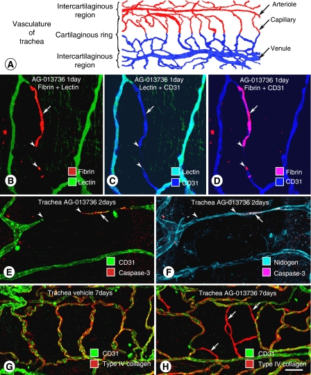
Figure 1.
Simple vascular network of tracheal mucosa used to examine effects of VEGF inhibition on normal blood vessels in adult mice. (A) Tracheal vasculature has a simple, repetitive network of arterioles, capillaries, and venules aligned with each cartilaginous ring (Baffert et al, 2004). (B–D) Confocal microscopic images of tracheal capillaries showing deposits of fibrin in nonpatent segment of tracheal capillary after inhibition of VEGF signalling by AG-013736 for 1 day. Fibrin deposit (arrow) is shown to be in a nonperfused capillary segment by absence of lectin binding, and is near a region of capillary regression that lacks CD31 immunoreactivity (arrowheads) (Baffert et al, 2006b). (E–F) Confocal images of tracheal vasculature showing apoptotic endothelial cells stained for activated caspase-3 (arrow), near region of capillary regression (arrowheads) shown by absence of CD31 immunoreactivity (E). Vascular basement membrane persists after endothelial cells regress, as shown by uninterrupted nidogen immunoreactivity (F) (Baffert et al, 2004). (G–H) Confocal micrographs show colocalisation of CD31 (green) and type IV collagen (red) on normal vasculature (G). After AG-013736 for 7 days, empty sleeves of type IV collagen (red, arrows) replace some normal mucosal blood vessels (H) (Inai et al, 2004). Scale bar in (H): 20 μm in (B–D); 25 μm in (E) and (F); 30 μm in (G) and (H).

