Figure 2.
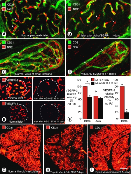
Regression of capillaries in vasculature of normal adult mice after inhibition of VEGF signalling. (A–D) Confocal microscopic images showing capillaries in pancreatic islets (A and B) and villi of small intestine (C and D) under baseline conditions and after VEGF inhibition. After Ad-sVEGFR-1 for 14 days, endothelial cells of some capillaries have regressed, leaving pericytes (red, NG2, arrowheads) at sites of regression (Kamba et al, 2006). (E) Comparison of VEGFR-2 and VEGFR-3 immunofluorescence in pancreatic islet capillaries after VEGF inhibition. Stronger endothelial cell VEGFR-2 immunoreactivity under baseline conditions (upper left) than after Ad-sVEGFR-1 for 14 days (upper right). Stronger endothelial cell VEGFR-3 immunoreactivity under baseline conditions (lower left) than after Ad-sVEGFR-1 for 14 days (lower right). (F) Bar graphs showing fluorescence intensities of VEGFR-2 and VEGFR-3 immunoreactivities under baseline conditions and after Ad-sVEGFR-1 for 14 days (Kamba et al, 2006). *P<0.05, significantly different from corresponding control. †P<0.05, significantly different from islets. (G–I) Fluorescence micrographs of thyroid capillaries stained for CD31 immunoreactivity show dense vascularity under baseline conditions (G), loss of half of the capillaries after AG-013736 for 7 days (H), and complete regrowth of vasculature during 14 days after end of treatment (I) (Kamba et al, 2006). Scale bar in I: 25 μm in (A–D); 40 μm in (E) and (F); 160 μm in (G–I).

