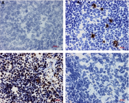
Figure 4.
Expression of p16 protein detected by immunohistochemistry. Immunohistochemistry was performed on frozen primary ESFT (n=37) and p16 protein detected using the CSA System. Tumours were negative (A), expressed in focal hotspots (B) or throughout the tumour (C). Homozygous deletion of CDKN2A correlated with loss of p16 protein expression in 3/4 tumours. Immunohistochemistry performed in the absence of primary antibody controlled for nonspecific binding of the primary antibody (D). Magnification × 400.

