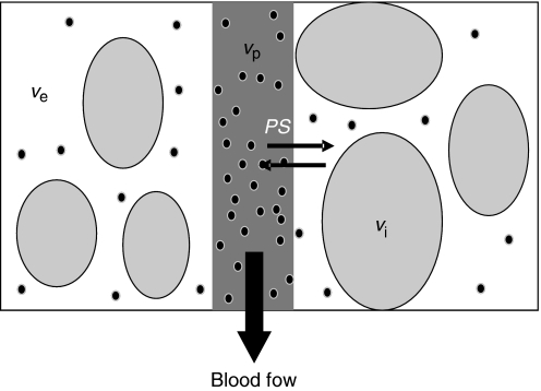
Figure 1.
Compartmental modelling of the tumour microvasculature: blood flows through the tumour enabling contrast media molecules (represented as black dots) to distribute in two potential compartments – the blood plasma volume vp and the volume of the extravascular extracellular space ve. Clinically available MRI contrast agents do not leak into the intracellular space vi. Contrast agent leakage is governed by the concentration difference between the plasma and the extracellular extravascular space and by the permeability and surface area of the capillary endothelia, expressed as PS.

