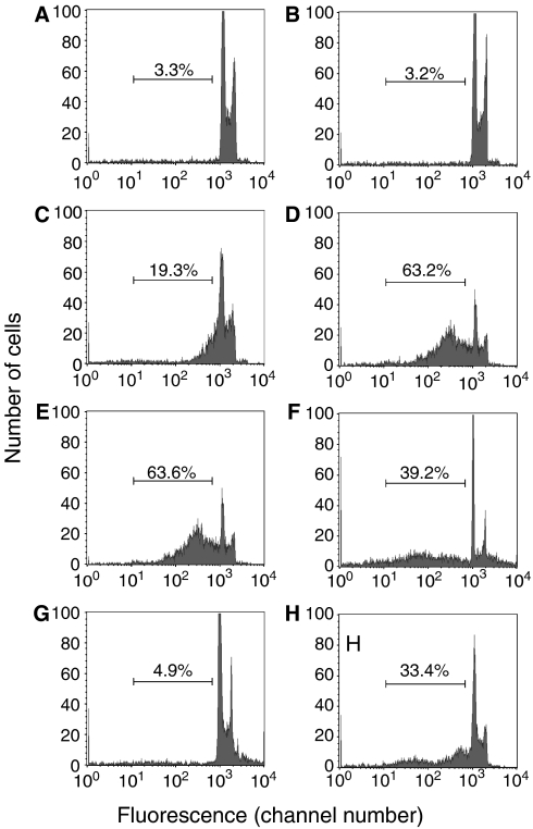
Figure 1.
Necrotic cells can have sub-G1 DNA content. Histograms of cells stained with propidium iodide, a DNA stain, are shown. (A) Control Jurkat cells. (B–E) Jurkat cells were killed by heating at 56°C and analyzed immediately after killing (B), or after incubation at 37°C for 24 h (C) or 48 h (D). (E) is the same as (D), except with a higher threshold setting, which excludes all cells smaller than the smallest normal cell. (F) Jurkat cells were killed by treatment with 0.005% digitonin for 5 min, then washed, and incubated for 6 h at room temperature. (G, H) Jurkat cells were killed by freeze–thawing and analyzed immediately after killing (G), or after incubation at 37°C for 3 days (H). The bar shows the region considered to represent cells with sub-G1 DNA content and the percentage of cells within this region is shown.

