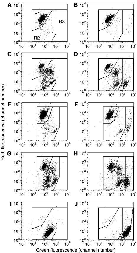
Figure 2.
Necrotic cells lose mitochondrial membrane potential (MMP), assayed by JC-1 staining, but can be distinguished from apoptotic cells by inclusion of Sytox green. Jurkat or Ramos cells were stained with JC-1 only (left graphs) or with JC-1 plus Sytox green (right graphs), which is a nuclear stain selective for dead cells. (A, B) Untreated Jurkat cells. (C, D) Jurkat cells treated overnight with 100 μM etoposide. Note that the apoptotic cells include two distinct populations, which can be considered early and late apoptotic cells based on the gradual loss of red staining. (E, F) Untreated Ramos cells. (G, H) Ramos cells treated with 20 nM paclitaxel overnight. (I, J) Jurkat cells killed by heating at 56°C for 45 min and examined immediately after killing. Regions R1, R2, and R3 indicated in (A) represent the normal, apoptotic, and lysed cells, respectively, after staining with JC-1 plus Sytox green. Note that these regions are slightly different, but similar for the two cells lines.

