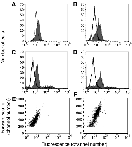
Figure 4.
Nonspecific staining of irradiated cells with fluorochrome-conjugated secondary Abs. Raji cells were irradiated with 10 Gy from a 137Cs irradiator, then washed and cultured in tissue culture medium for 1–3 days. At day 1 (A) or day 2 (B), cells were fixed and permeabilised, then stained by the procedure used for staining with anti-cleaved PARP (but without the primary Ab), using FITC-goat anti-mouse IgG. At day 2 (C) or day 3 (D) viable unfixed cells were stained with FITC-normal goat IgG. Results are shown for untreated cells (unfilled curve) and irradiated cells (filled curve). (E, F) Dot plots of forward scatter vs green fluorescence for the same two samples shown in (B): (E), control untreated cells; (F), cells 2 days after irradiation. Note that the irradiated cells are considerably larger and that fluorescence is correlated with cell size (as indicated by forward scatter).

