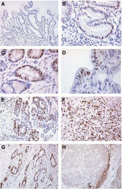
Figure 1.
Photomicrographs of HIF-1α immunohistochemistry in the gastric cancer progression sequence showing no staining in normal mucosa (A), weak nuclear staining in mucosal cells in H. pylori gastritis (B), moderate staining in intestinal metaplastia (C), distinct nuclear staining in high grade dysplasia (D), and strong staining in well (E) and poorly (F) differentiated intestinal adenocarcinoma (E) and revealing moderate HIF-1α staining; (D) High grade dysplasia showing distinct nuclear HIF-1α staining. Well (E) and poorly (F) differentiated intestinal adenocarcinoma showing distinct strong nuclear HIF-1α staining. Photomicrographs of HIF-1α immunohistochemistry in resected gastric cancer specimens showing focally positive (G) and invasive edge (H) patterns of staining.

